

 Joachim Zuther, Lymphedema Specialist.
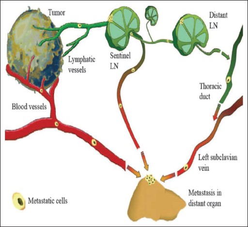
In metastasis, cancer cells break away from where they first formed (primary cancer), travel through the blood or lymph system and form new tumors, known as metastatic tumors in other parts of the body. The metastatic tumor is the same type of cancer as the primary tumor.
The lymphatic system consists of the circulating lymph fluid, a conducting network of lymphatic vessels, and lymphoid organs.
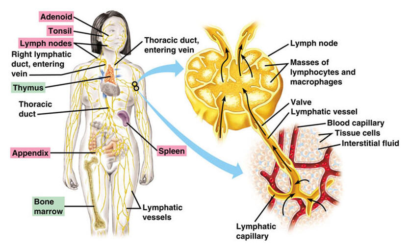
Lymphoid organs can be divided into two categories: Primary lymphoid organs (bone marrow and thymus), which are responsible for the production of T and B lymphocytes. T lymphocytes, or T-cells are a type of white blood cells (leukocytes); their roles include directly killing infected host cells and activating other immune cells. B lymphocytes, or B-cells, another sub type of leukocyte, are the triggers for immunity in the body fluids, providing defense against pathogens through different functions including the production of antibody molecules in body fluids.
The bone marrow is responsible for both the production of T lymphocytes and the creation and maturation of B lymphocytes, which will colonize the secondary lymphoid organs. B lymphocytes immediately join the circulatory system and travel to secondary lymphoid organs, whereas T lymphocytes travel from the bone marrow to the thymus, where they develop further.
Secondary lymphoid organs include the thymus, lymph nodes, spleen, tonsils, Peyer’s patches (located in the ileum region of the small intestine) and associated lymphoid tissue in mucous membranes. These peripheral lymphoid organs are arranged as a series of filters monitoring the contents of the lymph fluid, tissue fluid and blood. Secondary lymphoid tissues are also the location where mature lymphocytes are activated by antigens, thus initiating adaptive immune response.
Lymph Nodes
Lymph nodes are small anatomical structures that are an integral part of the immune system and filter lymph, the clear fluid that circulates through the lymphatic system. They drain lymph fluid from nearby organs or areas of the body and become swollen in response to infection and tumors. Clusters of lymph nodes are found in the neck, axilla (underarm), chest, abdomen, and groin. For example, there are about 20-40 lymph nodes in the axilla.
The number of these round, kidney or bean-shaped encapsulated lymphatic organs in an average human varies between 600-700, with most of the nodes located strategically in the intestines and the head-neck areas (place of entry for pathogens).
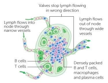
Lymph nodes have three main functions:
Protective function
They serve as filters for harmful material (cancer cells, pathogens, dust, dirt) in the lymph fluid
Immune function
Production of antigen-stimulated lymphocytes (antibodies). Lymphocytes are white blood cells, which circulate in the blood and the lymphatic system. They harbor in the lymph nodes and spleen and are part of the immune system responsible for both directly (T-cells and macrophages) and indirectly (B-cells producing antibodies) attacking foreign invaders. The antibodies produced in the lymph nodes leave the nodes via lymph collectors and travel within the lymph fluid to the blood for distribution throughout the body.
Thickening of the lymph fluid
Blood capillaries inside the lymph nodes reabsorb a large portion of the water content in the lymph fluid, thereby reducing the amount of lymph returning via the thoracic duct (and the right lymphatic duct) into the venous system.
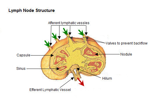
The size of lymph nodes in adults varies between 0.2 and 3cm. The shape, number and size of lymph nodes depend on factors such as age, sex and constitution. A set number of lymph nodes are present at birth; while lymph nodes increase and decrease in size during the course of life, they do not regenerate or vanish.
Most lymph nodes are embedded in fatty tissue and are arranged in either groups or chains. Their capsular area consists of dense connective tissue.
Lymph fluid enters the lymph nodes through lymph collectors (afferent lymph vessels), which perforate the capsular area, and leaves the nodes at an area known as the hilus via lymph collectors leading away from the nodes (efferent lymph vessels); lymph collectors also connect different lymph nodes with each other.
In order to ensure sufficient filter function, lymph fluid will pass in most cases through more than one lymph node before it re-enters the blood circulation.
Extensions of the capsular area of lymph nodes consist of connective tissue, called trabeculae, provide support for blood vessels entering into the nodes. These trabeculae compartmentalize the inside of the lymph node. A large number of lymphocytes and macrophages are located between the trabeculae and are connected by a loose net of fibers.
The lymph fluid circulates within the lymph node sinus system, which is located between the capsular area, the trabeculae and the clusters of defense cells. Upon entering the intra-nodal sinus system, the lymph flow is considerably slowed down, which enables defense cells to better identify and eliminate harmful substances.
In many cases, regional lymph nodes represent the first line of defense in the lymph transport. The drainage or tributary area of a group of regional lymph nodes may include several lymphatic territories. For example, the tributary area for the lymph nodes located in the groin (inguinal lymph nodes) consists of the legs, gluteal area, the external genitalia (skin), perineum, and the lower body quadrants (abdominal and lumbar areas). The tributary area for the axillary lymph nodes includes the arms and the upper body quadrants (chest and back).
How does Cancer appear, or spread to the Lymph Nodes?
Cancer can either start in lymph nodes, in which case it would be known as lymphoma, or it can spread there from somewhere else. When cancer cells break away from a primary tumor site, they can travel to other areas through either the bloodstream or the lymph system. If they travel through the lymph system, the cancer cells may end up in lymph nodes. Most of the escaped cancer cells die or are killed before they can start growing somewhere else; however, some might settle in a new area, begin to grow, and form new tumors. This spread of cancer to a new part of the body is called metastasis.
When cancer does spread to lymph nodes, it usually spreads to nodes near the tumor itself. These are the nodes that have been doing most of the work to filter out or kill the cancer cells.
The most common symptom of cancer in the lymph nodes is that one or more lymph nodes become swollen or feel hard, sometimes these nodes even become visible. The swelling or enlargement is called lymphadenopathy. If swollen lymph nodes are located inside the chest or deep in the abdominal area, the lymph nodes cannot be seen or felt. Often there are no symptoms. But sometimes swollen lymph nodes may press on nearby organs or structures, which can cause symptoms. In these cases, doctors may use scans or other imaging tests to look for enlarged nodes that are located deep in the body.
Secondary cancer in the lymph nodes may be diagnosed at the same time as the primary cancer. It may also be found during routine tests and scans after treatment. When cancer is present in a lymph node, a biopsy (samples of one or more nodes using needles) helps determine what type of cancer it is when the removed tissue or node is examined under a microscope. The cancer cells will look like the cancer cells of the tumor where they originated, so breast cancer cells in the lymphatic system will still look like breast cancer.
When a surgeon operates to remove a primary cancer, they may remove one or more of the nearby (regional) lymph nodes as well. Removal of part of one lymph node is known as a biopsy; when several lymph nodes are removed, it’s called lymph node dissection. When cancer has spread to lymph nodes, there is a higher risk that the cancer might come back after surgery. This information helps the physician decide whether more treatment (chemotherapy, immunotherapy, radiation), might be needed after the surgical removal.
What happens when Lymph Nodes are removed?
When lymph nodes are removed, it can leave the affected area without a way to drain away the lymph fluid. Many of the lymph vessels now run into a dead end where the node used to be, and fluid can back up. This may result in lymphedema, which can become a life-long problem. The higher the number of removed lymph nodes, the more likely lymphedema is to occur.
Lymphedema can be successfully treated and managed with Complete Decongestive Therapy (CDT).
Additional Reading: Metastatic cancer: What happens when cancer spreads?
Dear Lymphedema Blog Reader – if you like the contents on this website, please help to keep it going. A great amount of work and research is necessary to provide you with up-to-date information on this site. Your donation supports these efforts and associated administrative costs. Surplus funds will be donated to Lymphedema/Lipedema-related charitable endeavors. Please donate using the “Donate Now” button on the right upper hand of this page – Thank You!
Join Lymphedema Guru, a Facebook page solely dedicated to informing about all things related to lymphedema – news, support groups, treatment centers, and much more.


[…] How does Cancer appear, or spread to the Lymph Nodes? […]